ECR 2018 / C-2816

The Mesenteric Organ? - New concepts and a novel radiological perspective on its disease
Congress:
ECR 2018
Poster Number:
C-2816
Type:
Educational Exhibit
Keywords:
Abdomen, MR, CT, Education, Education and training, Mesentery, Peritoneum
Authors:
H. R. F. Dalla Pria1, F. Velloni1, R. A. Santiago1, M. S. Zacarias1, L. F. D. Silva1, F. Tamamoto1, A. C. Von Atzingen2, U. S. Torres1, G. D'Ippolito1; 1São Paulo/BR, 2Pouso Alegre/BR
DOI:
10.1594/ecr2018/C-2816

Table 1:
Table of contents

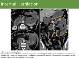
Fig. 11:
Left internal paraduodenal hernia

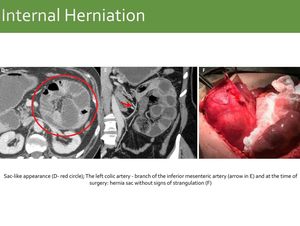
Fig. 12:
Same case as Fig. 11: Left internal paraduodenal hernia

Fig. 14:
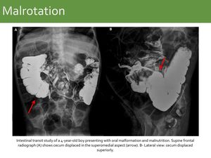
Malrotation: Case 1.

Fig. 15:
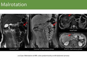
Malrotation Case 2.

Fig. 17:
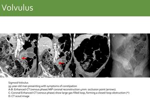
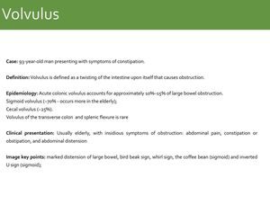
Sigmoid Volvulus

Fig. 20:
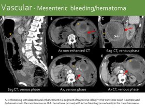
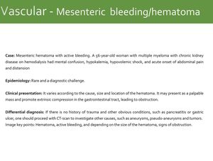
Vascular - Mesenteric bleeding/hematoma

Fig. 22:
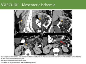
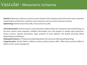
Vascular - Mesenteric ischemia

Fig. 24:
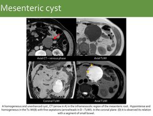
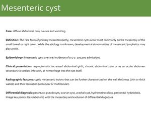
Mesenteric cyst

Fig. 26:
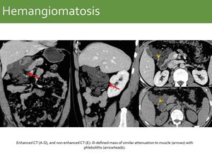
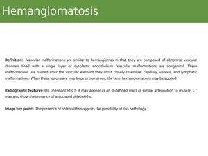
Hemangiomatosis

Fig. 28:
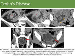
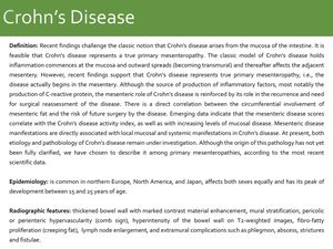
Crohn’s Disease

Fig. 30:
Mesothelioma

Fig. 31:
Same case as Fig. 30: Mesothelioma

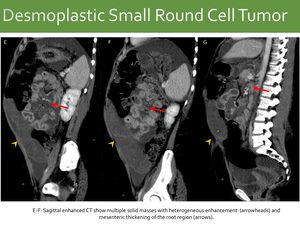
Fig. 33:
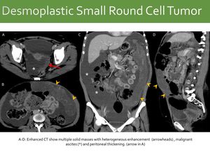
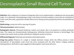
Desmoplastic Small Round Cell Tumor

Fig. 34:
Same case as Fig. 33: Desmoplastic Small Round Cell Tumor

Fig. 36:
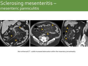
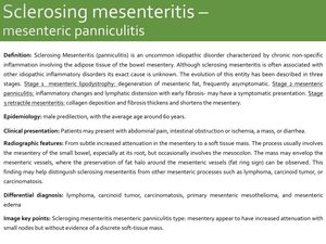
Sclerosing mesenteritis – mesenteric panniculitis

Fig. 38:
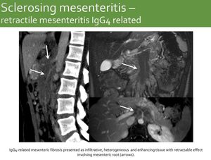
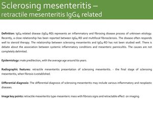
Sclerosing mesenteritis – Retractile mesenteritis IgG4 related

Fig. 40:
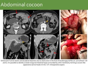
Abdominal cocoon

Fig. 42:
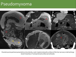
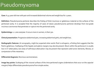
Pseudomyxoma

Fig. 44:
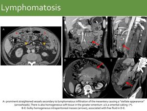
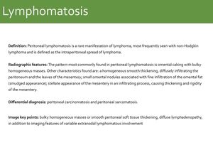
Lymphomatosis

Fig. 46:
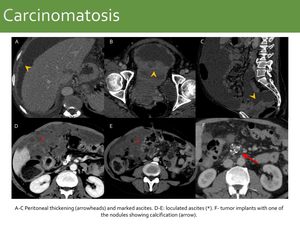
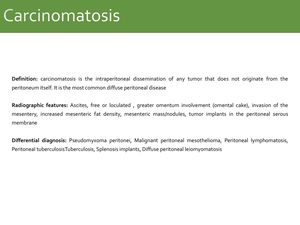
Carcinomatosis

Fig. 48:
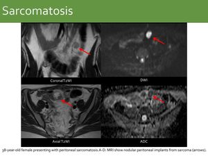
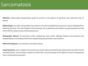
Sarcomatosis

Fig. 50:
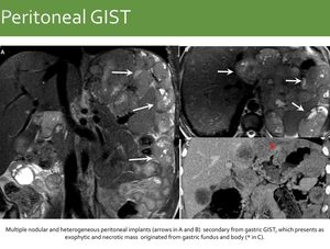
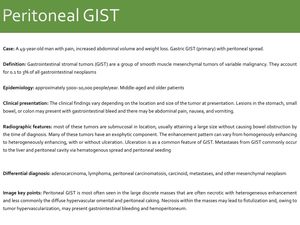
Peritoneal GIST

Fig. 52:
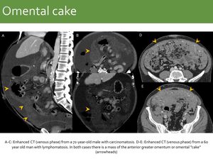
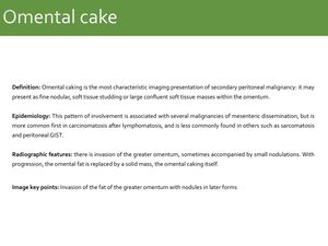
Omental cake

Fig. 54:
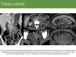
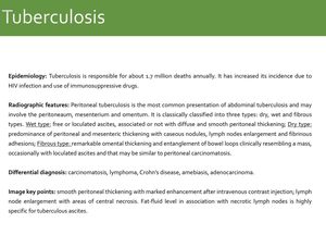
Tuberculosis

Fig. 56:
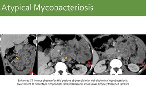
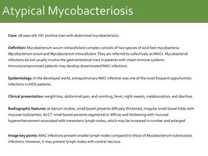
Atypical Mycobacteriosis

Fig. 58:
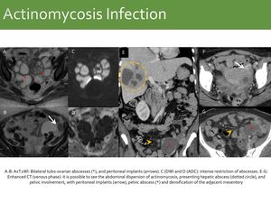
Actinomycosis Infection

Fig. 60:
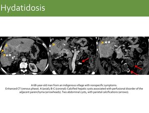
Hydatidosis

Fig. 62:
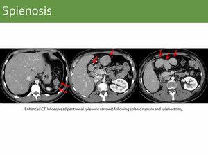
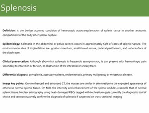
Splenosis

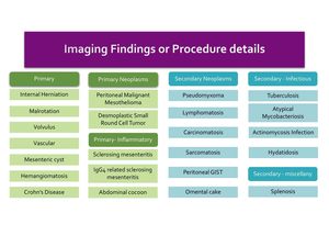
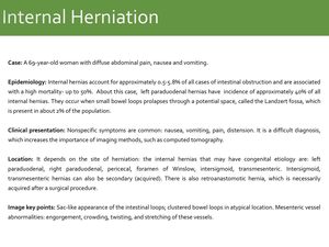
Fig. 13:
Internal Herniation

Fig. 16:
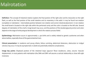
Malrotation

Fig. 19:
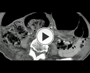
Volvulus

Fig. 21:
Vascular - Mesenteric bleeding/hematoma

Fig. 23:
Vascular - Mesenteric ischemia

Fig. 25:
Mesenteric cyst

Fig. 27:
Hemangiomatosis

Fig. 29:
Crohn’s Disease

Fig. 32:
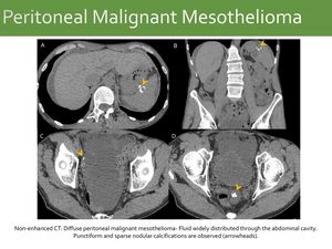
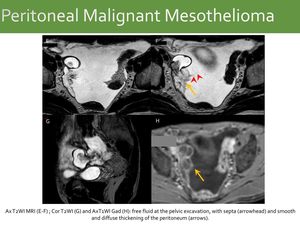
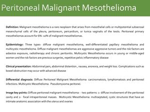
Peritoneal Malignant Mesothelioma

Fig. 35:
Desmoplastic Small Round Cell Tumor

Fig. 37:
Sclerosing mesenteritis – mesenteric panniculitis

Fig. 39:
Sclerosing mesenteritis – retractile mesenteritis IgG4 related

Fig. 41:
Abdominal cocoon

Fig. 43:
Pseudomyxoma

Fig. 45:
Lymphomatosis

Fig. 47:
Carcinomatosis

Fig. 49:
Sarcomatosis

Fig. 51:
Peritoneal GIST

Fig. 53:
Omental cake

Fig. 55:
Tuberculosis